

COSMETIC:
(888) 803-3762
MEDICAL:
(519) 836-8558

22
Sep
Mole Mapping in Guelph: Early Melanoma Detection
TL;DR: Mole mapping combines total-body photography with sequential digital dermoscopy so new or changing moles get caught early. In the strongest cohort study, 53.3% of melanomas found this way were still in situ, meaning caught before any invasion into deeper skin. Depth drives outcomes: melanomas 0.8 mm or thinner showed 93.4% ten-year melanoma-specific survival. However, mole mapping is a surveillance tool for higher-risk people, not a treatment, and screening average-risk adults without symptoms remains unproven. If you have many moles, atypical moles, or a personal or family history of melanoma, structured surveillance is where the evidence points.
The Canadian Cancer Society estimates 11,300 Canadians will be diagnosed with melanoma in 2026, 6,200 men and 5,100 women, and that 1,250 will die of it (Canadian Cancer Society, 2026 estimates). In my practice in Guelph, the melanoma stories that end well share one feature: the lesion was found early, often as a subtle change nobody would’ve flagged without a systematic way to look. That’s the entire logic of mole mapping. I’m Dr. Dusan Sajic, a dermatologist here in Guelph. In this guide I’ll cover what mole mapping involves, what the evidence genuinely shows, who benefits, and where the honest limits sit. Because millimetres and months decide a great deal in this disease.
Key Takeaways
- Canada expects 11,300 melanoma diagnoses and 1,250 deaths in 2026 (Canadian Cancer Society, 2026 estimates).
- Digital follow-up of 618 high-risk patients found 98 melanomas, 53.3% still in situ, with invasive tumours at a median of just 0.5 mm (Salerni et al., JAAD, 2012).
- Melanomas 0.8 mm or thinner carried 93.4% ten-year melanoma-specific survival (Annals of Surgical Oncology, 2018).
- Dermatologists detected melanoma with 76.9% sensitivity versus 37.5% for general practitioners, and dermoscopy raised detection odds 13.3-fold (JAMA Dermatology analysis, 2024).
What Is Mole Mapping and How Does It Work?
Mole mapping is a structured skin-cancer surveillance method that pairs standardized total-body photographs with sequential digital dermoscopy: magnified imaging of individual moles, repeated over time. The goal is simple: catch new or changing lesions early by comparing today’s skin against a fixed baseline instead of memory. Who needs it? Mainly higher-risk people: those with many moles (roughly 100 or more), multiple atypical moles, or a personal or family history of melanoma, not average-risk adults. For that group, the evidence is strong. In a ten-year study of 618 high-risk patients, this two-step method found 98 melanomas, and 53.3% were still in situ, meaning caught before invading beyond the skin’s top layer (Salerni et al., JAAD, 2012). In my Guelph practice, it’s the backbone of high-risk follow-up.
The invasive melanomas in that cohort were caught at a median Breslow thickness, the depth of invasion measured in millimetres, of just 0.5 mm. Consequently, the real message isn’t that photography finds more moles. It’s that structured comparison finds the same cancers earlier, when they’re thinnest and most treatable.
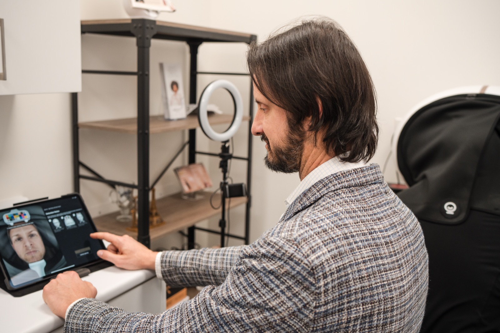
Here’s what mole mapping looks like inside our medical dermatology practice. Total-body photography is the baseline step: standardized photographs of your skin surface that every later visit gets compared against. We add dermoscopic images of the moles worth tracking, then schedule re-imaging. Change between visits, a brand-new lesion, growth, or a shift in colour, is what triggers a closer look or a biopsy. Meanwhile, stability is evidence too. In fact, documented stability can spare a harmless-but-odd-looking mole an unnecessary excision.
Why Do Millimetres Matter in Melanoma?
Because outcomes track tumour depth remarkably closely. Melanomas 0.8 mm thick or thinner showed 93.4% ten-year and 85.7% twenty-year melanoma-specific survival in a large surgical series (Annals of Surgical Oncology, 2018). What I tell patients is simple: catch a melanoma while it’s thin, and the long-term outlook is strongly favourable.
Meanwhile, Canadian numbers frame the same point from the other side. Five-year net survival for melanoma in Canada is 90% overall (Canadian Cancer Society / Statistics Canada, 2019-2021 data). Canada doesn’t publish stage-specific survival, so the Canadian Cancer Society cites international figures instead. In the AJCC series, five-year melanoma-specific survival runs 99% at Stage 1A versus 50% at Stage 4 (Canadian Cancer Society, citing international data). A 49-point spread between earliest and latest stage. That’s the entire argument for early detection, in one line.
Can one millimetre honestly matter that much? Patients ask me that all the time, and it can. Thin lesions usually mean simpler surgery and better odds; thicker ones mean staging workups and harder conversations.
Does Mole Mapping Actually Catch Melanoma Earlier?
The evidence leans yes, with caveats I’ll give you straight. In fact, across 14 studies covering 12,082 patients, melanomas found under photographic monitoring were thinner at diagnosis: median 0.30-0.50 mm, versus 0.48-0.83 mm in groups without photography (Hornung et al., IJERPH systematic review, 2021).
Now the caveats, because you deserve them. These are observational studies, not randomized trials, and the review’s authors flagged genuine risks of bias and uneven study quality. Meanwhile, people who enrol in surveillance may also differ from people who don’t. However, the direction of the finding is consistent across studies, and it matches the high in-situ detection rates seen in dedicated follow-up cohorts. One more honest limit: no surveillance method catches everything. For instance, a fast-growing lesion can appear between visits, so a new or changing spot always warrants a call rather than waiting for your next mapping session.
Who Actually Needs Mole Mapping?
Not everyone; risk concentrates sharply, and surveillance should follow it. How many moles put you in the high-risk group? People with 101 to 120 common moles carry 6.89 times the melanoma risk of people with very few (95% CI 4.63-10.25). In fact, having five atypical (dysplastic) moles, moles with irregular features under magnification, multiplies risk 6.36-fold (Gandini et al., European Journal of Cancer meta-analysis, 2005).
In our Guelph clinic, the groups I most often move into structured surveillance are:
- Many moles, roughly 100 or more; I see this weekly, and nobody can reliably track that much change from memory
- Multiple atypical moles; a 2024 risk analysis put their odds ratio at 3.01 (British Journal of Dermatology, 2024)
- A previous melanoma: risk of a second primary runs about nine times the general population’s, with roughly 7.6% cumulative risk within five years (second-primary melanoma study)
- Melanoma in a close relative, which carried an odds ratio of 1.79 in the same 2024 analysis (British Journal of Dermatology, 2024)
That said, if none of those describe you, periodic skin exams and good self-checks may serve you well. That’s not me talking anyone out of care; it’s matching the tool to the risk.
Doesn’t the USPSTF Say Skin Screening Isn’t Proven?
For average-risk adults, yes, and patients deserve that context. In 2023 the US Preventive Services Task Force assigned an “I” grade to visual skin screening of asymptomatic, average-risk adults: insufficient evidence to weigh benefits against harms (USPSTF, 2023; a US recommendation).
Here’s the catch: the fine print matters. That statement explicitly does not cover people with a personal or family history of melanoma, people with symptomatic or changing lesions, or people already under high-risk surveillance. Those exclusions describe precisely the patients mole mapping exists for. Consequently, my position matches the evidence on both sides: I don’t push whole-population screening, and I do recommend structured surveillance once a risk assessment justifies it. Unsure which side of that line you’re on? That’s exactly what an assessment settles, and I’ve written about when to see a dermatologist in Guelph if you want the practical signals.
How Should You Check Your Own Moles Between Visits?
Monthly, using the ABCDE rule, and with realistic expectations about what self-checks can do. In a 2024 analysis, dermatologists detected melanoma with 76.9% sensitivity on clinical examination versus 37.5% for general practitioners (Chen et al., JAMA Dermatology, 2024). Dermoscopy is a polarized magnification tool; in the same analysis, adding it raised detection odds 13.3-fold. Self-checks raise flags; trained eyes and instruments confirm them.
The checklist itself has history: the ABCD criteria were introduced in 1985, and E was added in 2004 (Abbasi et al., JAMA, 2004). As defined by the American Academy of Dermatology, a US organization (AAD):
- A – Asymmetry: one half doesn’t match the other
- B – Border: irregular, scalloped, or poorly defined edges
- C – Colour: varied shades of brown or black, sometimes patches of red, white, or blue
- D – Diameter: larger than about 6 mm, though melanomas can be smaller
- E – Evolving: changing in size, shape, or colour
Which letter matters most? E is the one I weight most heavily. A stable, boring mole is rarely the enemy. The changing one is, and change is exactly what mapping is built to expose.
Mole Mapping FAQs
How often should mole mapping be repeated?
It depends on your risk profile, and we set the interval at your assessment. For instance, the cohort that caught 53.3% of melanomas in situ used scheduled follow-up imaging over ten years (Salerni et al., JAAD, 2012). In my practice, yearly re-mapping is typical, with closer checks for individual moles we’re actively watching.
How is mole mapping different from a regular skin check?
Documentation and magnification. A regular exam leans on memory; mapping compares today’s skin against baseline images, and monitored melanomas trend thinner at diagnosis, median 0.30-0.50 mm versus 0.48-0.83 mm without photography (IJERPH systematic review, 2021). Dermoscopy alone raised detection odds 13.3-fold (JAMA Dermatology, 2024).
Can mole mapping prevent melanoma?
No, and I won’t claim it can. It’s early detection, not prevention: the payoff is catching tumours thin, where 0.8 mm or less carried 93.4% ten-year melanoma-specific survival (Annals of Surgical Oncology, 2018). Meanwhile, prevention still rests on sun protection; finding disease early is what mapping does.
I’ve had melanoma before. Do I actually need this?
Your risk justifies structured follow-up more than almost anyone’s. After a first melanoma, the risk of a second primary is roughly nine times the general population’s, with about 7.6% cumulative risk within five years (second-primary melanoma study). Consequently, ongoing surveillance is standard advice I give every melanoma survivor.
What happens if mapping finds something suspicious?
Usually a dermoscopic reassessment, then a small biopsy if concern holds; many flagged moles turn out benign. That said, acting early is the whole point: international AJCC data show five-year melanoma-specific survival of 99% at Stage 1A versus 50% at Stage 4 (Canadian Cancer Society, citing international data).
Book a Mole Mapping Assessment in Guelph
If you carry higher risk, many moles, atypical moles, or a personal or family history of melanoma, don’t leave surveillance to memory; in my experience, memory is exactly where subtle change hides. Request a consultation at deRMA Skin Institute. We’ll assess your risk honestly, tell you whether mole mapping is genuinely indicated for you, and set the right follow-up rhythm if it is.
Medically reviewed by Dr. Dusan Sajic, MD, PhD — dermatologist, deRMA Skin Institute, Guelph, ON.
Sources
1. Canadian Cancer Society. Melanoma skin cancer statistics (2026 estimates). https://cancer.ca/en/cancer-information/cancer-types/melanoma-skin/statistics 2. Canadian Cancer Society. Survival statistics for melanoma skin cancer (Canadian net survival; international AJCC stage data). https://cancer.ca/en/cancer-information/cancer-types/melanoma-skin/prognosis-and-survival/survival-statistics 3. Salerni G et al. Benefits of total-body photography and sequential digital dermoscopy (“two-step method of digital follow-up”) in high-risk patients. Journal of the American Academy of Dermatology, 2012. https://pubmed.ncbi.nlm.nih.gov/21683472/ 4. Long-term survival of patients with thin (T1) cutaneous melanomas (0.8 mm cut point). Annals of Surgical Oncology, 2018. https://pubmed.ncbi.nlm.nih.gov/29330716/ 5. Hornung A et al. The value of total body photography for the early detection of melanoma: a systematic review. International Journal of Environmental Research and Public Health, 2021. https://pmc.ncbi.nlm.nih.gov/articles/PMC7916771/ 6. Chen et al. Melanoma detection sensitivity by clinician type and dermoscopy. JAMA Dermatology, 2024. https://pmc.ncbi.nlm.nih.gov/articles/PMC11561728/ 7. Gandini S et al. Meta-analysis of risk factors for cutaneous melanoma: I. Common and atypical naevi. European Journal of Cancer, 2005. https://pubmed.ncbi.nlm.nih.gov/15617989/ 8. Risk of a second primary melanoma after a first primary melanoma. https://pubmed.ncbi.nlm.nih.gov/20231496/ 9. Melanoma risk factors and prediction analysis (family history, atypical naevi). British Journal of Dermatology, 2024. https://academic.oup.com/bjd/article/190/2/174/7240713 10. US Preventive Services Task Force. Skin Cancer: Screening (Grade I statement), 2023. https://www.uspreventiveservicestaskforce.org/uspstf/recommendation/skin-cancer-screening 11. Abbasi NR et al. Early diagnosis of cutaneous melanoma: revisiting the ABCD criteria (E for Evolving added). JAMA, 2004. https://jamanetwork.com/journals/jamadermatology/fullarticle/478587 12. American Academy of Dermatology. The ABCDEs of melanoma. https://www.aad.org/public/diseases/skin-cancer/find/at-risk/abcdes
Dusan Sajic, MD, PhD
Richard Backstein, MD

Sonja Sajic, CCPA

Toni Alberto, CCPA
With more than 20 years of experience, deRMA Skin Institute strive to offer patients the most advanced treatments available to keep their skin healthy and looking its best. Board Certified Dermatologist, Dusan Sajic, MD, PhD, board-certified Plastic and Reconstructive Surgeon, Richard Backstein, MD, FRCSC, Sonja Sajic, CCPA, and Toni Alberto CCPA are committed to providing state-of-the-art medical, surgical and cosmetic treatments to all patients in Guelph, Cambridge, Kitchener, Hamilton, Milton, and surrounding areas.
Contact Us
Fields marked * are required
If you are an existing patient inquiring about appointments and services, please contact
Guelph Cosmetic Department: (888) 803-3762 cosmetic@dermaskininstitute.com
Guelph Medical Department: (519) 836-8558 patientcare@dermaskininstitute.com